
Kidney stones smaller than 5 mm have the best chance of passing naturally, while stones approaching or exceeding 10 mm are much more likely to require medical treatment. However, a kidney stone size chart provides only an estimate. The stone’s location, shape, urinary obstruction, infection, pain level, and your kidney function can significantly change the outcome.
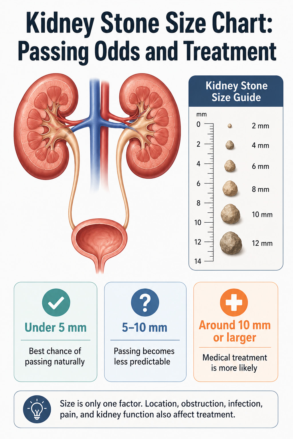
Most published passing rates apply to ureteral stones—stones that have left the kidney and entered the tube connecting the kidney to the bladder. A small stone that remains inside the kidney may stay there without symptoms and should not be assigned the same passing odds.
Quick Kidney Stone Size Chart
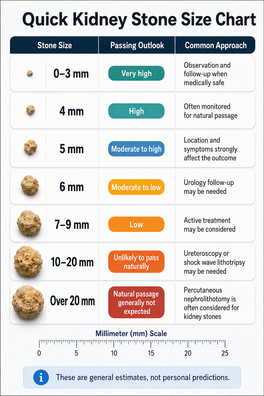
The following chart provides a practical overview of how stone size may affect natural passage and treatment decisions.
| Kidney stone size | General chance of passing | Common medical approach |
|---|---|---|
| 0–3 mm | Very high | Observation, symptom control, and follow-up when medically safe |
| 4 mm | High | Often monitored for natural passage |
| 5 mm | Moderate to high | Observation may be appropriate; treatment depends heavily on location and symptoms |
| 6 mm | Moderate to low | May pass, but urology follow-up and possible treatment are more likely |
| 7–9 mm | Low | Active treatment is commonly considered |
| 10–20 mm | Unlikely to pass naturally | Ureteroscopy or shock wave lithotripsy is often needed |
| Over 20 mm | Natural passage is generally not expected | Percutaneous nephrolithotomy is usually considered for stones inside the kidney |
These categories are not guarantees. A 5 mm stone near the bladder may pass more easily than a 4 mm stone lodged near the kidney or associated with significant swelling.
The 2026 European Association of Urology guidelines report that almost 75% of ureteral stones smaller than 5 mm pass spontaneously. The passage rate was about 62% when all stones measuring 5 mm or larger were grouped together, although the likelihood continued to decrease as stone size increased.
Kidney Stone Passing Odds by Millimeter
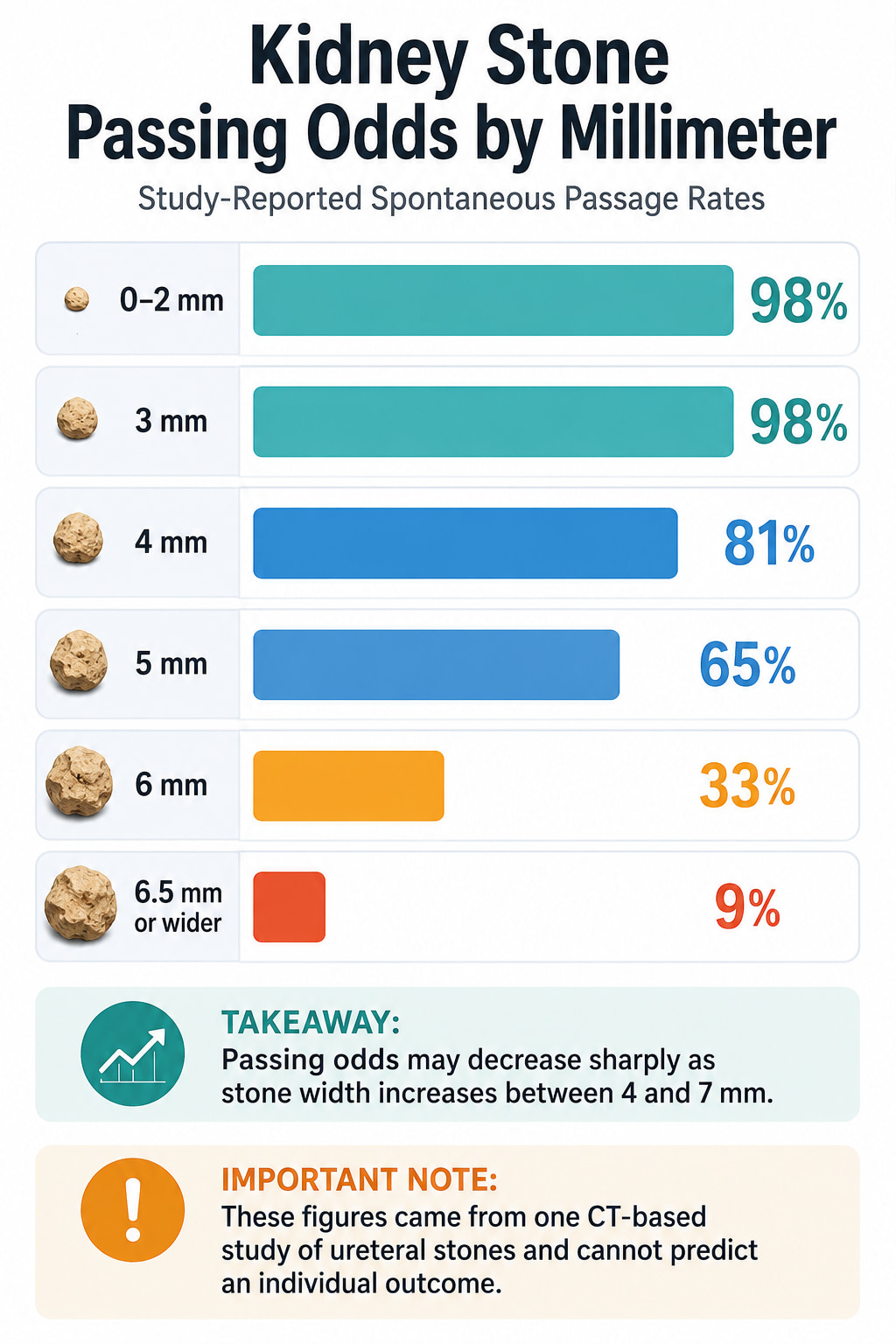
One CT-based study followed 392 ureteral stones and reported the following spontaneous passage rates over 20 weeks.
| Stone width on CT | Spontaneous passage rate |
|---|---|
| 0–2 mm | 98% |
| 3 mm | 98% |
| 4 mm | 81% |
| 5 mm | 65% |
| 6 mm | 33% |
| 6.5 mm or wider | 9% |
These percentages came from one retrospective study and should not be treated as a personal prediction. The estimates were also based on the stone’s width on CT, which may not be the same measurement that appears first on every imaging report.
The sharp decline between 5 and 7 mm helps explain why urologists become more likely to discuss a procedure as a stone grows. Still, some larger stones pass, and some very small stones become stuck.
What Size Kidney Stone Can Pass Naturally?
A ureteral stone under 5 mm is generally the most likely to pass without a procedure. Stones measuring 5–10 mm may also pass, but the chance becomes progressively lower as the stone gets wider.
A stone may be suitable for observation when:
- Pain can be controlled
- There are no signs of infection
- Urine can pass around the stone
- Kidney function remains stable
- The stone has a reasonable likelihood of passing
- The patient can return for follow-up imaging and evaluation
Observation does not mean ignoring the stone. A clinician may repeat imaging to confirm that it has passed and is no longer blocking urine flow.
Can a 5 mm Kidney Stone Pass?
Yes. A 5 mm ureteral stone can pass naturally.
The CT study above reported a 65% passage rate for stones measuring exactly 5 mm. That percentage should be interpreted cautiously because a stone’s location and the patient’s anatomy can raise or lower the chance.
A distal 5 mm stone close to the bladder generally has a better outlook than one in the upper ureter.
Can a 6 mm Kidney Stone Pass?
A 6 mm stone can pass, but the probability is substantially lower than it is for a 4 or 5 mm stone.
In the same CT-based study, approximately 33% of 6 mm ureteral stones passed spontaneously within the observation period. A clinician may still recommend a monitored trial of passage when there is no infection, dangerous obstruction, uncontrolled pain, or kidney impairment.
Can a 7 mm Kidney Stone Pass?
It is possible for a 7 mm stone to pass, especially when it is located near the bladder. However, passage is less likely, and the stone has a greater risk of remaining lodged in the ureter.
A urologist may discuss ureteroscopy, shock wave lithotripsy, or a carefully monitored trial of passage based on the stone’s exact position and the patient’s condition.
Can a 10 mm Kidney Stone Pass?
A 10 mm stone is unlikely to pass naturally. Although spontaneous passage is not impossible, relying on it may prolong pain or obstruction.
Stones around this size generally require prompt urologic evaluation. Treatment is often recommended when the stone is blocking urine, causing persistent symptoms, or unlikely to move on its own.
Why Stone Location Changes the Passing Odds
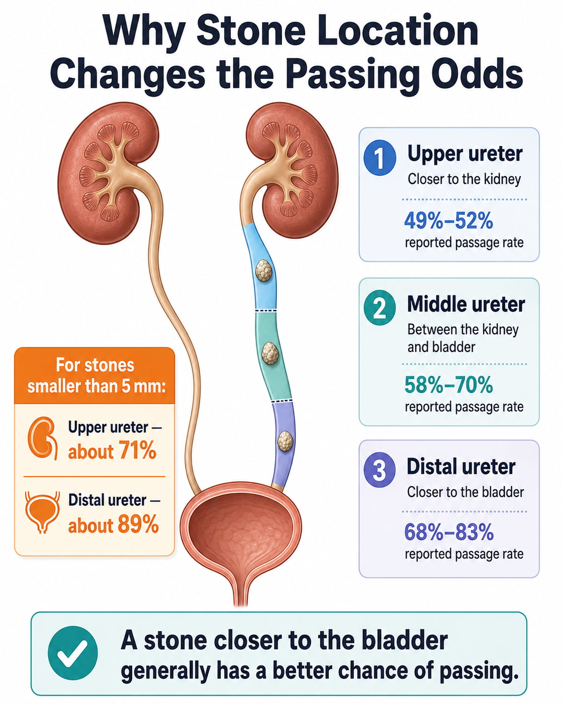
Kidney stones generally become painful after they move from the kidney into the ureter. The ureter is a narrow muscular tube that carries urine to the bladder.
Doctors commonly describe ureteral stone locations as:
- Upper or proximal ureter: Closer to the kidney
- Middle ureter: Between the kidney and bladder
- Lower or distal ureter: Closer to the bladder
The EAU reports the following spontaneous passage ranges by location:
| Stone location | Reported spontaneous passage rate |
|---|---|
| Upper ureter | 49%–52% |
| Middle ureter | 58%–70% |
| Distal ureter | 68%–83% |
For stones smaller than 5 mm, the guideline estimates an 89% passage chance when the stone is in the distal ureter, compared with 71% when it is in the upper ureter.
These figures show why two people with stones of the same size may receive different recommendations.
Stones Inside the Kidney Are Different
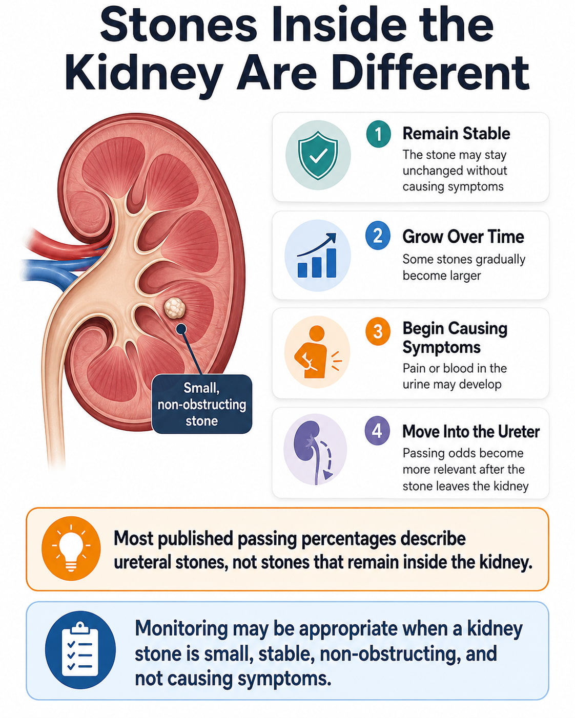
Passing charts are less useful for stones that remain inside the kidney. A non-obstructing kidney stone may:
- Remain unchanged
- Gradually grow
- Begin causing pain or blood in the urine
- Move into the ureter
- Eventually require treatment
The natural history of small, asymptomatic kidney stones is unpredictable. In studies of active surveillance, spontaneous passage ranged from 3% to 29%, stone growth from 5% to 66%, symptom development from 7% to 77%, and eventual surgical intervention from 7% to 26%.
A stable kidney stone under 10 mm may sometimes be monitored with periodic imaging. Treatment becomes more appropriate if the stone grows, causes symptoms, creates an obstruction, contributes to infection, or presents a special risk based on the patient’s health or circumstances.
How Long Does a Kidney Stone Take to Pass?
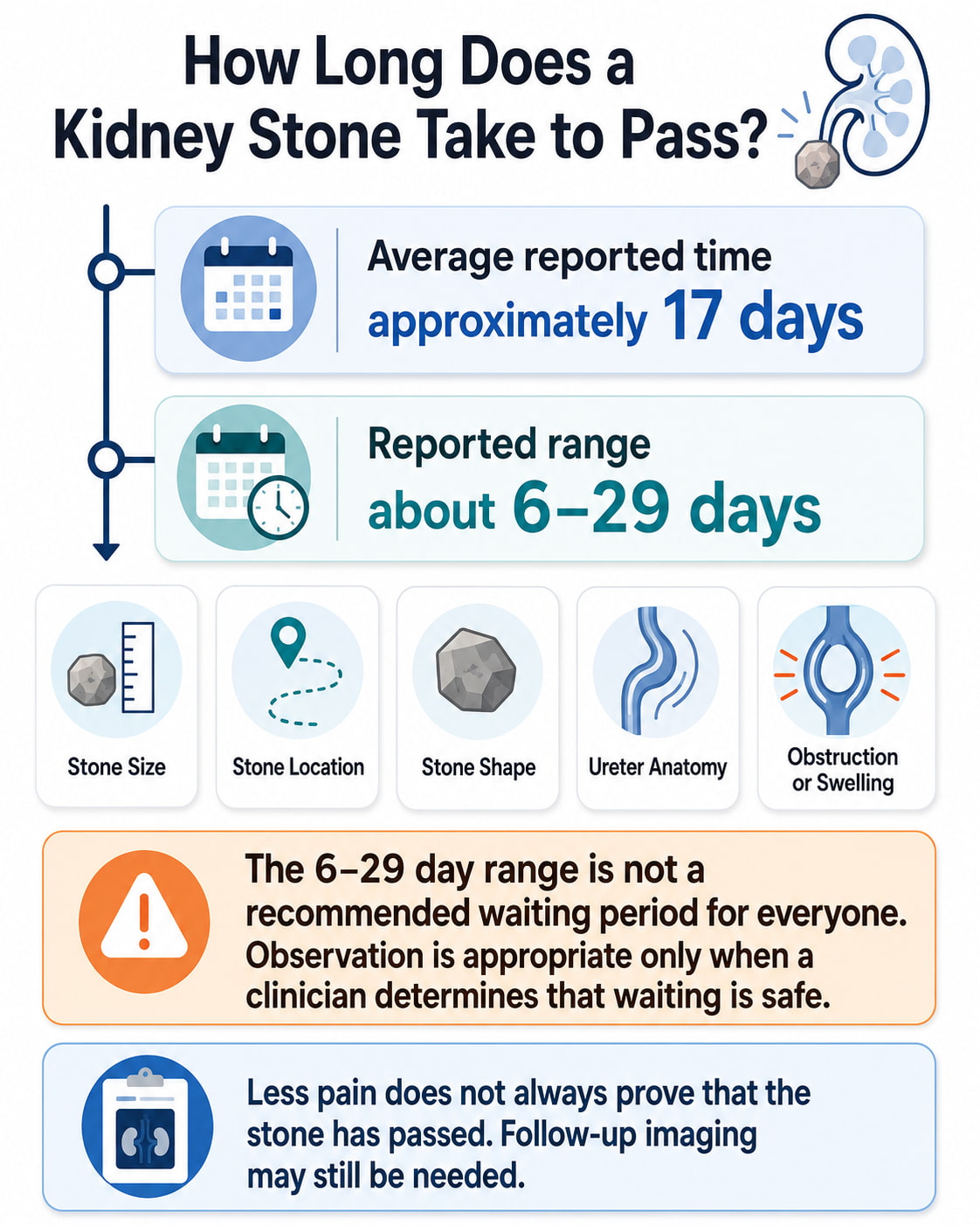
The average expulsion time reported in the EAU guideline is approximately 17 days, with a range of about 6–29 days.
That does not mean every patient should wait 29 days. Waiting is appropriate only when a clinician determines that observation is safe.
Passage time depends on:
- Stone size
- Stone location
- Stone shape
- Ureter anatomy
- Swelling or obstruction
- Whether the stone is moving
- Previous stone history
- Medical expulsive therapy
Pain may come and go as the ureter contracts around the stone. A reduction in pain does not always prove that the stone has passed. Follow-up imaging may be needed when the stone was not visibly collected.
Kidney Stone Treatment by Size
Treatment cannot be selected from size alone. Doctors also consider stone location, density, composition, symptoms, infection risk, urinary anatomy, kidney function, and whether the stone is causing hydronephrosis.
General Treatment Chart
| Stone type and size | Possible approach |
|---|---|
| Small ureteral stone under 5 mm | Observation and periodic evaluation when no complications are present |
| Distal ureteral stone 5–10 mm | Observation, prescription medical expulsive therapy, ureteroscopy, or shock wave lithotripsy |
| Ureteral stone 10–20 mm | Ureteroscopy is commonly preferred; shock wave lithotripsy may be appropriate in selected cases |
| Kidney stone under 10 mm | Monitoring if asymptomatic; shock wave lithotripsy or ureteroscopy if treatment is needed |
| Kidney stone 10–20 mm | Shock wave lithotripsy, ureteroscopy, or selected use of percutaneous treatment |
| Kidney stone over 20 mm | Percutaneous nephrolithotomy is generally the first-line treatment |
The UK’s National Institute for Health and Care Excellence recommends shock wave lithotripsy or ureteroscopy for many smaller renal and ureteral stones, ureteroscopy for most 10–20 mm ureteral stones, and percutaneous nephrolithotomy for renal stones larger than 20 mm. Individual practice may differ based on local expertise and patient-specific factors.
Observation and Follow-Up
Observation may be offered for a newly diagnosed small ureteral stone when active removal is not immediately necessary.
During observation, the care plan may include:
- Pain control
- Instructions about fluid intake
- Urine straining to collect the stone
- Monitoring for fever or worsening symptoms
- Follow-up imaging
- A scheduled urology appointment
Observation should be stopped if infection, uncontrollable pain, persistent obstruction, or worsening kidney function develops.
Medical Expulsive Therapy
Medical expulsive therapy uses prescription medication to support the passage of a ureteral stone.
The updated EAU guideline recommends offering an alpha-blocker as one possible treatment for distal ureteral stones measuring approximately 5–10 mm. The expected benefit is greatest for stones larger than 5 mm that are close to the bladder. This use is off-label and should be discussed with a clinician.
Medical expulsive therapy is not appropriate when urgent stone removal or drainage is necessary. It should be discontinued and the patient reassessed if infection, uncontrolled pain, or declining kidney function develops.
Shock Wave Lithotripsy
Shock wave lithotripsy uses externally generated shock waves to break a stone into smaller pieces. The fragments then pass through the urinary tract.
Advantages may include:
- No incision
- Outpatient treatment in many cases
- Lower complication rates than ureteroscopy in some comparisons
Limitations include:
- More than one session may be required
- Some hard or dense stones do not break easily
- Lower-pole kidney stones may be harder to clear
- Stone fragments can temporarily block the ureter
Shock wave treatment is often considered for smaller kidney or ureteral stones when the stone can be accurately targeted.
Ureteroscopy
During ureteroscopy, a urologist passes a thin scope through the urethra and bladder and into the ureter or kidney. The stone can then be removed with a small basket or fragmented with a laser.
According to the National Institute of Diabetes and Digestive and Kidney Diseases, ureteroscopy is performed with anesthesia, and many patients can return home on the same day. A temporary ureteral stent may be placed to support urine flow after the procedure.
Ureteroscopy generally offers a better chance of becoming stone-free after one procedure than shock wave lithotripsy. However, it is more invasive and may have a higher complication rate.
Percutaneous Nephrolithotomy
Percutaneous nephrolithotomy, or PCNL, is commonly used for large kidney stones.
During PCNL, the surgeon reaches the kidney through a small opening in the back and removes or breaks up the stone. It is generally recommended as the first treatment for kidney stones larger than 20 mm because less invasive approaches may require several sessions and leave a greater amount of stone behind.
PCNL normally requires anesthesia and may involve a hospital stay.
When Does a Kidney Stone Need Treatment?
A stone does not need to reach a specific size before treatment becomes necessary. Even a small stone may require intervention if it causes a serious complication.
The EAU lists the following reasons for removing a ureteral stone:
- A low likelihood of natural passage
- Persistent pain despite appropriate medication
- Continued urinary obstruction
- Reduced kidney function
- Obstruction of both ureters
- Obstruction affecting a solitary functioning kidney
Kidney stones that remain inside the kidney may need treatment when they grow, cause pain or blood in the urine, block urine flow, contribute to infection, or create special risks for the patient.
When Is a Kidney Stone an Emergency?
An obstructed kidney combined with infection or an inability to produce urine is a urologic emergency. Urgent drainage may be required with a ureteral stent or a nephrostomy tube. Definitive stone removal is generally delayed until the infection has been controlled.
Seek urgent medical care for:
- Fever or chills with flank, abdominal, or groin pain
- Inability to urinate or passing only a very small amount
- Severe pain that does not improve with prescribed medication
- Repeated vomiting or inability to keep liquids down
- Cloudy or foul-smelling urine with pain or fever
- Increasing weakness, confusion, or feeling severely unwell
- Stone symptoms in someone with one functioning kidney
- A known obstructing stone with worsening symptoms
The NIDDK kidney stone symptom guide includes fever, chills, vomiting, painful urination, reduced urine output, and cloudy or bad-smelling urine among symptoms that require medical assessment.
What to Do While Waiting for a Stone to Pass
Follow the plan provided by your healthcare professional. The appropriate approach may differ for someone with kidney disease, heart failure, pregnancy, a solitary kidney, or another medical condition.
Practical steps may include:
- Take only the pain medicine recommended for you.
- Drink liquids according to your clinician’s instructions.
- Use a urine strainer if asked so the stone can be analyzed.
- Record pain, fever, vomiting, and changes in urine output.
- Attend follow-up imaging even if the pain improves.
- Contact your clinician if symptoms worsen or the stone does not pass as expected.
Do not begin an alpha-blocker, antibiotic, potassium citrate, or another kidney stone medication without medical guidance.
Frequently Asked Questions
Is a 4 mm kidney stone considered large?
No. A 4 mm stone is considered small and has a relatively high chance of passing if it is already in the ureter. The CT-based study cited above reported an 81% spontaneous passage rate for 4 mm stones, although location and obstruction still matter.
What is the largest kidney stone that can pass naturally?
There is no absolute maximum. Some stones larger than 7 mm pass, while some stones smaller than 5 mm become stuck. However, natural passage becomes increasingly unlikely as the stone approaches 10 mm.
Does a lower ureteral stone pass more easily?
Generally, yes. Distal ureteral stones located close to the bladder have higher passage rates than stones close to the kidney. The EAU reports passage ranges of 68%–83% for distal stones compared with 49%–52% for upper ureteral stones.
Should every kidney stone be removed?
No. Some small, non-obstructing kidney stones can be monitored when they are stable and not causing symptoms. Treatment may become necessary if the stone grows, causes pain, contributes to infection, blocks urine, or affects kidney function.
Can drinking more water make a large stone pass?
Adequate fluid intake supports normal urine production, but it cannot guarantee that a large or obstructing stone will pass. A stone that is too wide, firmly impacted, or causing complications may need a procedure.
Can kidney stones be dissolved with medicine?
Some uric acid stones may be dissolved by medically raising the urine pH. Most calcium-based stones cannot simply be dissolved with medication and may need to pass naturally or be removed. The stone type must be properly evaluated before dissolution therapy is considered.
Can pain stop before the stone has passed?
Yes. Pain may decrease when the stone stops moving, moves to a less sensitive area, or reaches the bladder. It may also remain lodged without causing continuous pain. Follow-up imaging may be needed to confirm passage and rule out silent obstruction.
Conclusion
A kidney stone size chart can help estimate whether a stone is likely to pass, but it cannot replace an individual medical evaluation. Stones under 5 mm generally have the best passing odds. Stones measuring 5–10 mm have less predictable outcomes, while stones around 10 mm or larger are increasingly likely to need a procedure.
The safest next step is to confirm the stone’s exact size and location, check for urinary obstruction or infection, and follow the monitoring or treatment plan recommended by a healthcare professional. Seek urgent care for fever, chills, inability to urinate, uncontrolled pain, repeated vomiting, or signs of serious illness.
This content is for informational purposes only and not medical advice.
References
- European Association of Urology: Urolithiasis Guidelines, 2026
- National Institute of Diabetes and Digestive and Kidney Diseases: Kidney Stone Treatment
- National Institute of Diabetes and Digestive and Kidney Diseases: Kidney Stone Symptoms and Causes
- National Institute for Health and Care Excellence: Renal and Ureteric Stone Recommendations
- PubMed: Size Matters—Ureteral Stone Width, Location, and Spontaneous Passage