
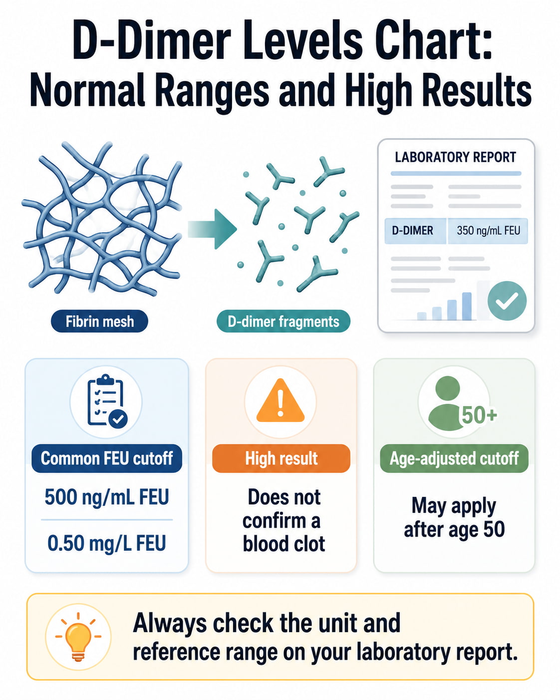
A common adult D-dimer cutoff is 500 ng/mL FEU, equal to 0.50 mg/L FEU, but the correct normal range is the reference limit printed on your laboratory report. A result above that limit is considered positive or elevated. However, it does not prove that you have a deep vein thrombosis, pulmonary embolism, or another blood clot.
D-dimer testing is most useful for ruling out a clot in people whose symptoms and medical history suggest a low or intermediate clinical probability. Your age, pregnancy status, reporting units, medications, recent surgery, and other health conditions can all affect how the result should be interpreted.
D-Dimer Levels Chart at a Glance
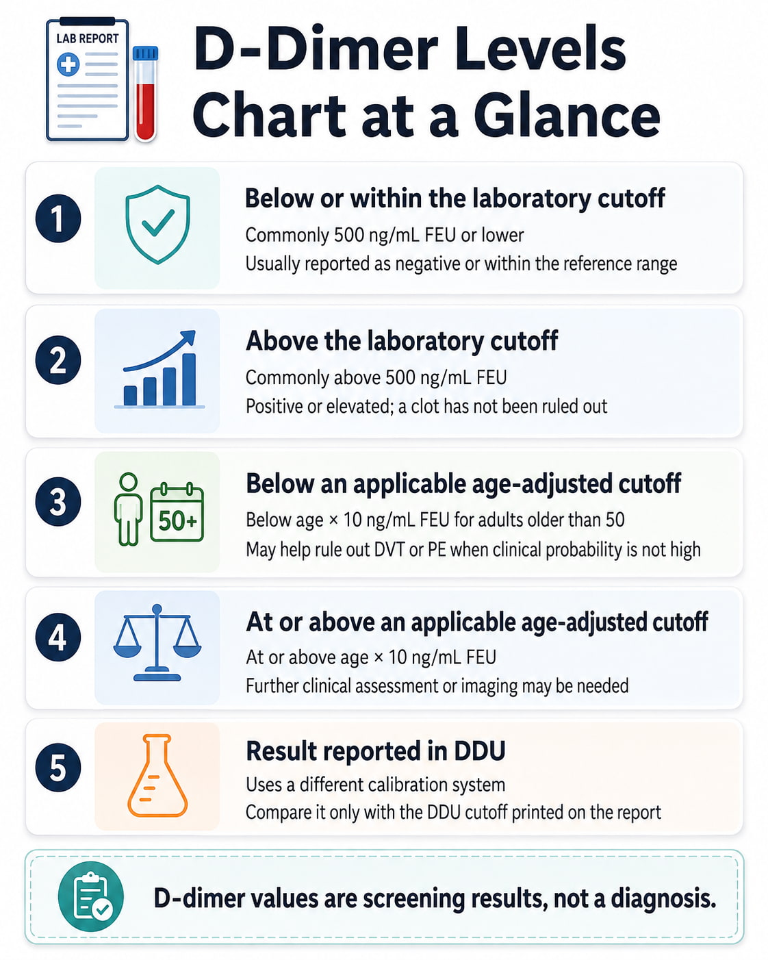
| D-dimer result | Common FEU value | General meaning |
|---|---|---|
| Below or within the laboratory cutoff | Commonly 500 ng/mL FEU or lower | Usually reported as negative or within the reference range |
| Above the laboratory cutoff | Commonly above 500 ng/mL FEU | Positive or elevated; a clot has not been ruled out |
| Below an applicable age-adjusted cutoff | Below age × 10 ng/mL FEU for adults older than 50 | May help rule out DVT or PE when clinical probability is not high |
| At or above an applicable age-adjusted cutoff | At or above age × 10 ng/mL FEU | Further clinical assessment or imaging may be needed |
| Result reported in DDU | Uses a different calibration system | Must be compared with the DDU cutoff printed on the report |
These categories are general guides rather than universal diagnostic ranges. For one widely used assay, Mayo Clinic Laboratories lists 500 ng/mL FEU or lower as its reference value. Other laboratories may use different assays, units, and cutoff wording.
What Is a D-Dimer Test?
D-dimer is a protein fragment produced when the body breaks down cross-linked fibrin, an important part of a blood clot. Small amounts may be present during normal clot formation and breakdown, while higher concentrations indicate increased activity in the clotting and clot-dissolving systems.
A D-dimer blood test may be used when a healthcare professional is evaluating possible:
- Deep vein thrombosis, or DVT
- Pulmonary embolism, or PE
- Disseminated intravascular coagulation, or DIC
- Other conditions involving increased clot formation and breakdown
The test does not show where a clot is located. It also cannot determine the clot’s size or severity.
What Is a Normal D-Dimer Level?
For many FEU assays, a D-dimer result around 500 ng/mL FEU or lower is considered within the reference range. Some laboratories use “below 500,” while others include 500 as a negative result.
The exact cutoff depends on the assay. Always check these three details on your report:
- The numerical result
- The reporting unit
- The laboratory reference limit
A result cannot be interpreted safely from the number alone.
Common D-Dimer FEU Unit Equivalents
The following values represent the same FEU concentration:
| Reporting unit | Equivalent value |
|---|---|
| ng/mL FEU | 500 ng/mL |
| µg/L FEU | 500 µg/L |
| mg/L FEU | 0.50 mg/L |
| µg/mL FEU | 0.50 µg/mL |
For example, 0.72 mg/L FEU is the same concentration as 720 ng/mL FEU.
These conversions apply only when both values use FEU. They should not be used to convert an FEU result into DDU.
FEU vs. DDU: Why the Units Matter
D-dimer tests use two main calibration systems.
| Reporting system | Full name | Common fixed cutoff |
|---|---|---|
| FEU | Fibrinogen equivalent units | Often near 500 ng/mL FEU |
| DDU | D-dimer units | Often near 230–250 ng/mL DDU, depending on the assay |
An FEU number is generally about twice the corresponding DDU number, but the relationship is not exact for every test system. A result of 400 ng/mL FEU and a result of 400 ng/mL DDU therefore do not mean the same thing.
The American Society of Hematology advises using the laboratory’s own methodology, units, and cutoff instead of manually converting between FEU and DDU.
How to Read the Units on Your Report
Look for wording such as:
- ng/mL FEU
- µg/L FEU
- mg/L FEU
- ng/mL DDU
- µg/L DDU
Do not compare a DDU result with an online chart labeled FEU. When the unit is unclear, ask the laboratory or ordering clinician which cutoff applies.
Age-Adjusted D-Dimer Levels Chart
D-dimer commonly increases with age, even when an acute blood clot is not present. For adults older than 50, clinicians may use an age-adjusted cutoff rather than the fixed 500 ng/mL FEU threshold.
The commonly used formula is:
Age-adjusted D-dimer cutoff = age × 10 ng/mL FEU
Because ng/mL and µg/L have the same numerical value, the formula may also be written as age × 10 µg/L FEU.
| Age | Common age-adjusted cutoff |
|---|---|
| 50 or younger | 500 ng/mL FEU |
| 55 | 550 ng/mL FEU |
| 60 | 600 ng/mL FEU |
| 65 | 650 ng/mL FEU |
| 70 | 700 ng/mL FEU |
| 75 | 750 ng/mL FEU |
| 80 | 800 ng/mL FEU |
| 85 | 850 ng/mL FEU |
| 90 | 900 ng/mL FEU |
For example, the age-adjusted cutoff for a 72-year-old would be:
72 × 10 = 720 ng/mL FEU
A result must generally be below the applicable threshold to be considered negative under that strategy.
When Is an Age-Adjusted Cutoff Used?
The age-adjusted calculation is not intended for everyone. It is usually applied when:
- The patient is older than 50.
- A highly sensitive FEU assay is used.
- Clinical assessment suggests a low or intermediate probability of a clot.
- The patient is being evaluated through a validated diagnostic pathway.
The 2026 American Heart Association and American College of Cardiology pulmonary embolism guideline states that an age-adjusted D-dimer below the threshold can effectively exclude PE and avoid imaging in adults with low or intermediate clinical probability. It should not be used to dismiss concerning symptoms in someone with a high clinical probability of PE.
2026 Research Update on Suspected DVT
Age adjustment has long been used when evaluating possible pulmonary embolism. Newer evidence also supports carefully selected use for suspected leg DVT.
The 2026 ADJUST-DVT study included 3,205 emergency department outpatients. Among patients whose clinical probability was not high, using the age-adjusted cutoff identified additional people in whom DVT could be ruled out without imaging. In participants aged 75 or older, the percentage with a negative D-dimer increased from 8.7% with the fixed cutoff to 26.1% with age adjustment, with no false-negative results identified in that study subgroup.
These findings apply to the study’s structured diagnostic pathway. They do not mean that adults should calculate a cutoff and rule out DVT without professional assessment. (JAMA ADJUST-DVT study)
What Does a High D-Dimer Result Mean?
A high D-dimer means the amount of fibrin breakdown material in the blood is above the assay’s cutoff. It shows that clot formation and breakdown may be occurring somewhere in the body, but it does not identify the reason.
A positive result may lead to additional testing when a person has symptoms or risk factors for DVT or PE. It should not be treated as a stand-alone diagnosis.
Examples of Result Interpretation
Example 1: 420 ng/mL FEU
If the laboratory cutoff is 500 ng/mL FEU, this would generally be considered negative. It is most reassuring when the clinical probability of DVT or PE is low or intermediate.
Example 2: 680 ng/mL FEU in a 40-year-old
This is above the common fixed cutoff. Age adjustment would not normally apply because the person is younger than 50. The result does not confirm a clot, but further evaluation may be appropriate.
Example 3: 680 ng/mL FEU in a 70-year-old
The common age-adjusted threshold would be 700 ng/mL FEU. The result may be considered below the age-adjusted cutoff when a clinician has determined that the patient is suitable for that diagnostic strategy.
Example 4: 400 ng/mL DDU
This cannot be interpreted using a 500 ng/mL FEU chart. It must be compared with the laboratory’s DDU reference limit.
Causes of High D-Dimer Levels
Blood clots are important causes of elevated D-dimer, but many other conditions can increase the result.
Clot-Related Causes
- Deep vein thrombosis
- Pulmonary embolism
- Disseminated intravascular coagulation
- Other active thromboembolic conditions
Other Possible Causes
- Recent surgery
- Physical trauma
- A large bruise or hematoma
- Active or recent bleeding
- Pregnancy
- Infection
- Systemic inflammation
- Cancer
- Liver disease
- Heart disease
- Rheumatoid arthritis
- Older age
- Prolonged immobility
- Recent hospitalization
An elevated result may also occur when several of these factors are present at the same time. This is why D-dimer has high sensitivity for detecting increased clotting activity but relatively low specificity for identifying one particular disease.
Can D-Dimer Be High Without a Blood Clot?
Yes. A high D-dimer does not automatically mean that DVT or PE is present.
For example, someone recovering from surgery may have an elevated result because surgery activates clotting and tissue-repair processes. Pregnancy, infection, inflammation, cancer, aging, and limited mobility can also produce positive results without an acute DVT or PE.
The test is therefore usually more useful when it is negative in an appropriately selected patient than when it is positive. A positive result tells the clinician that a clot may still need to be investigated.
Is There a Dangerous D-Dimer Level?
There is no universally accepted D-dimer number that automatically means a blood clot is dangerous, large, or life-threatening.
Online charts sometimes divide elevated results into categories such as mildly high, moderately high, severely high, or critical. These are not standardized diagnostic classifications.
Very high values can occur with serious conditions, including major blood clots, severe infection, cancer, extensive trauma, or DIC. However, the number alone cannot determine:
- Whether a clot is present
- Where a clot is located
- How large the clot is
- Whether the condition is immediately life-threatening
- What treatment is required
Mayo Clinic Laboratories notes that the degree of D-dimer elevation does not definitively correlate with the severity of the associated condition. Symptoms, vital signs, medical history, clinical probability, and imaging are more important than assigning a “danger level” to the number.
How Healthcare Professionals Use D-Dimer
A healthcare professional does not usually interpret D-dimer in isolation. The test is often combined with a clinical probability assessment.
Low or Intermediate Clinical Probability
A negative D-dimer may allow DVT or PE to be ruled out without imaging, depending on the assay and diagnostic pathway.
High Clinical Probability
A D-dimer test may not be sufficient. The clinician may proceed directly to imaging because a normal result should not override a strongly concerning clinical presentation.
Positive D-Dimer
A positive result does not confirm a clot. It indicates that further evaluation may be needed, particularly when symptoms or risk factors are present.
Clinical decision tools may consider factors such as recent surgery, cancer, previous blood clots, rapid heart rate, one-sided leg swelling, coughing up blood, and whether another diagnosis is more likely.
What Tests May Be Ordered After a High Result?
The next step depends on the suspected location of a clot and the person’s overall condition.
Doppler or Compression Ultrasound
An ultrasound may be used to examine the deep veins of the legs or arms for DVT.
CT Pulmonary Angiography
CT pulmonary angiography uses contrast material to look for a clot in the pulmonary arteries. It is commonly used when PE is suspected and the person can safely receive contrast.
Ventilation-Perfusion Scan
A ventilation-perfusion, or V/Q, scan compares airflow and blood flow in the lungs. It may be considered when CT angiography is not suitable.
According to MedlinePlus, imaging such as Doppler ultrasound, CT angiography, or a V/Q scan may be ordered after an abnormal result. The appropriate test depends on symptoms, clinical probability, pregnancy status, kidney function, allergies, and available resources.
Do not start aspirin or a prescription blood thinner based only on a D-dimer number. These medicines can cause serious bleeding and should be used only under appropriate medical guidance.
D-Dimer Levels During Pregnancy
D-dimer commonly rises during pregnancy, especially later in pregnancy. As a result, more pregnant patients test above the standard fixed cutoff even when pulmonary embolism is not present.
There is no single trimester-specific D-dimer range that can be safely applied to every pregnant patient because:
- Assays and units differ.
- Normal values vary between individuals.
- D-dimer frequently rises as pregnancy progresses.
- Symptoms of pregnancy and PE can overlap.
- The result must be combined with a pregnancy-specific clinical pathway.
The 2026 AHA/ACC guideline states that pregnancy-adapted YEARS criteria may be reasonable for identifying pregnant adults who do not require imaging for suspected PE. This is a clinician-directed decision strategy, not a self-calculation based on D-dimer alone.
Can a Normal D-Dimer Miss a Blood Clot?
A normal D-dimer is reassuring only when used in the correct clinical setting. It should not be used as the sole reason to dismiss severe or strongly suggestive symptoms.
Blood-thinning medicine may lower D-dimer and contribute to a false-negative result. Assay performance and the person’s clinical probability also matter. A healthcare professional may order imaging despite a normal result when the likelihood of DVT or PE remains high.
When to Seek Urgent Medical Care
Do not wait for a D-dimer test result when symptoms suggest pulmonary embolism or another emergency.
Possible Symptoms of DVT
Contact a healthcare professional promptly for:
- New swelling in one leg or arm
- Leg pain or tenderness
- Warm skin over the affected area
- Red, darkened, or discolored skin
- Unexplained one-sided limb swelling
Possible Symptoms of Pulmonary Embolism
Seek immediate emergency medical care for:
- Sudden shortness of breath
- Chest pain, especially pain that worsens with breathing
- Rapid breathing
- A fast or irregular heartbeat
- Coughing up blood
- Fainting, severe dizziness, or marked weakness
- Sudden unexplained low oxygen levels
Both DVT and PE require timely evaluation. Pulmonary embolism can be life-threatening, and a person with possible PE symptoms should not delay care while trying to interpret a laboratory number.
Frequently Asked Questions
Is 500 ng/mL a high D-dimer?
It depends on the assay and wording of the laboratory reference range. For many FEU assays, 500 ng/mL is the upper reference limit. Some laboratories consider 500 negative, while others define a negative result as below 500. Check the cutoff printed beside your result.
What D-dimer level confirms a blood clot?
No D-dimer level confirms DVT or PE by itself. Even a very high result can have several possible causes. Imaging and clinical assessment are required to confirm the diagnosis and locate a clot.
What is the normal D-dimer level in mg/L?
A common FEU cutoff is 0.50 mg/L FEU, which is equivalent to 500 ng/mL FEU. A DDU result uses a different cutoff and should not be compared directly with this value.
How is the age-adjusted D-dimer calculated?
For an adult older than 50, the common FEU formula is age multiplied by 10 ng/mL. For example, the cutoff for an 80-year-old would be 800 ng/mL FEU. The calculation is used only within an appropriate clinical pathway.
Why is my D-dimer high if imaging found no clot?
Possible reasons include recent surgery, trauma, pregnancy, inflammation, infection, cancer, liver disease, heart disease, older age, bleeding, or immobility. The result may remain nonspecific even after a clot has been excluded.
Can blood-thinning medicine affect D-dimer?
Yes. Anticoagulant medicine may lower D-dimer and contribute to a false-negative result. Tell the clinician whether you have already taken heparin, warfarin, apixaban, rivaroxaban, or another anticoagulant.
Should an elevated D-dimer be repeated?
Repeating the test is not always the appropriate next step. When an acute clot is suspected, clinical assessment and imaging may be more useful than waiting for another blood test. Follow the instructions of the clinician who ordered the test.
Conclusion
A D-dimer levels chart can help you understand the basic reference cutoff, but the number must always be read with its units, laboratory range, age, symptoms, and clinical context. A common adult cutoff is 500 ng/mL FEU, while adults older than 50 may qualify for an age-adjusted threshold under a clinician-directed diagnostic strategy.
A negative result can help rule out DVT or PE in appropriately selected patients. A high result means that further evaluation may be necessary, not that a dangerous blood clot has already been diagnosed. Contact the ordering healthcare professional for personalized interpretation, and seek immediate care for sudden breathing difficulty, chest pain, fainting, or coughing up blood.
This content is for informational purposes only and not medical advice.